
A fun animation about the development of tamariki and rangatahi brains by the NZ Classification Office in collaboration with youth health expert Dame Sue Bagshaw.
Source: New Zealand Classification Office
transcribeTranscript
[Music]
Our brains change just as much as our bodies as we grow.
That's why we understand films, tv and media differently at different ages.
When we're born, our brain has all the basic structures in place, but because we're so new lots of parts aren't yet fully switched on to transmit messages.
At this stage the base of our brain is pretty much running the show.
It's responsible for controlling things like our bodily functions and our fear response.
But as we learn to do things like eat, walk and talk, different parts of our brain power up at different times.
Like the cortex which is responsible for our ability to think.
While it develops we can't do a lot of complex thinking and reasoning.
That's why children can find it hard to tell the difference between fantasy and reality.
It's why they can think what they see in films or on tv is real.
As we grow our limbic system develops.
This part of the brain lays down memories and processes emotions.
And so does our amygdala, which recognises danger.
It links all our feelings up into the cortex where we can start to make sense of those feelings and what they mean.
When we're in our teens the amygdala is calling the shots.
That's why the teenage years can be an emotional rollercoaster.
The link up to the cortex is in the process of being switched on and some parts are working well and some not.
It's also the reason that teens are more prone to risk-taking and scaring themselves with things like horror films.
They also get a reward from enjoying the thrills.
At this age our amygdala is also more developed than our cortex.
That's why young people still need help thinking critically about what they're watching.
And that can continue right into their 20s as our brains don't fully develop until then.
At any age though, even as fully grown adults, it's good for all of us to think carefully and critically about what we're watching.
For more information visit classificationoffice.govt.nz.
Key points about adolescent brain development
- a safe environment where rangatahi (young people) have consistent, loving support is vital for the brain to develop well
- when you are talking to rangatahi, be careful to check what emotion they are seeing in you
- always acknowledge your child's emotions first and then help them to think about what they're feeling
- rangatahi need adults to believe in them and encourage them
- rangatahi respond better to rewards than to punishment
- rangatahi need clear, consistent boundaries
- it's important that parents respect their child's growing capacity and independence
When brain development happens
The brain develops very rapidly in the first 3 to 5 years of life. All the structures and building blocks are present by the age of 9. The different centres of the brain develop and become functionally connected over time. The last part to mature is the prefrontal lobe. This happens during adolescence.
Many things affect brain development, including genes, individual and environmental factors.
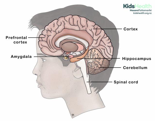
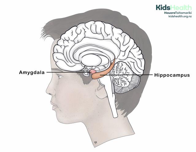
This illustration shows a side view of the brain in a young person, with key areas of the brain labelled.
Source: KidsHealth
transcribeTranscript
This illustration shows a side-view drawing of a young person’s head with the brain shown in cross-section. The following brain areas are coloured and labelled:
- Prefrontal cortex (front of the brain)
- Amygdala (small, deep area near the centre)
- Hippocampus (curved area in the centre of the brain)
- Cortex (outer layer of the brain)
- Cerebellum (rounded area at the lower back of the brain)
- Spinal cord (stem running down from the brain)
The labels are connected to each brain area with straight black lines.
At the top right is the KidsHealth logo with the website: kidshealth.org.nz.
Why the brain takes so long to develop
Human beings are the only animals that are born completely helpless. We also have the biggest size of adult brain. It is believed that if we were born with an adult-sized brain, our heads would not fit through our mothers' hips. The brain keeps developing after birth. This helps us adapt to our living environment - whether it be a city or a rainforest. Being able to adapt gives us a better chance of surviving.
We used to think that when growth stops after puberty, development was complete. Then, scientists invented MRI scanners. MRI scanners showed the brain continues to change for a long time after puberty has finished. In fact, brain development may not be complete until around 30 years of age.
The following image shows that the brain doesn't change much in size between 5 and 20 years of age. What changes is the colour. The blue shows connections happening between the parts of the brain that are already formed.
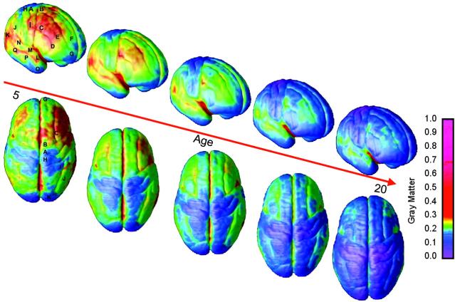
This illustration shows brain scans from ages 5 to 20. The colours show how it develops and matures over time.
Source: Copyright 2004 - National Academy of Sciences, U.S.A.
transcribeTranscript
A series of 10 brain scan images arranged in two rows, showing brain development from age 5 to 20. Each column shows two views of the same brain:
- The top row shows the right-side (lateral) view
- The bottom row shows the top-down view
A red diagonal arrow across the image is labelled 'Age', with 5 at the left end and 20 at the right.
Each brain is coloured using a heatmap that represents the amount of grey matter (GM) across the brain’s surface. The colour bar on the right shows GM volume, with red and yellow representing higher grey matter and blue/purple representing lower grey matter.
This image sequence shows how the brain changes from age 5 to 20. While the size of the brain stays similar, the colours shift from red/yellow/green to blue—showing how the brain matures over time. This process is called grey matter maturation.
How the brain develops
Some people like to think of the brain in 4 parts.
1. The spinal cord and the base of the brain
Delivers messages to and from all parts of the body.
Controls what happens in the parts you don't have to consciously think about. This includes the heart, lungs and stomach.
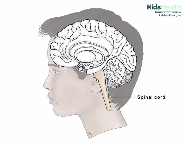
This illustration shows the adolescent brain in cross-section with the spinal cord highlighted.
Source: KidsHealth
transcribeTranscript
A side-view illustration of a young person’s head, with the brain shown in cross-section. The spinal cord is highlighted in light yellow and runs down from the base of the brain into the neck and spine. A black line points to it, with a label that says 'Spinal cord'.
The rest of the brain is shown in greyscale with detailed folds and internal structures, including the cerebellum.
At the top right is the KidsHealth logo with the website: kidshealth.org.nz.
2. The cerebellum
Controls and coordinates movement and other brain processes.
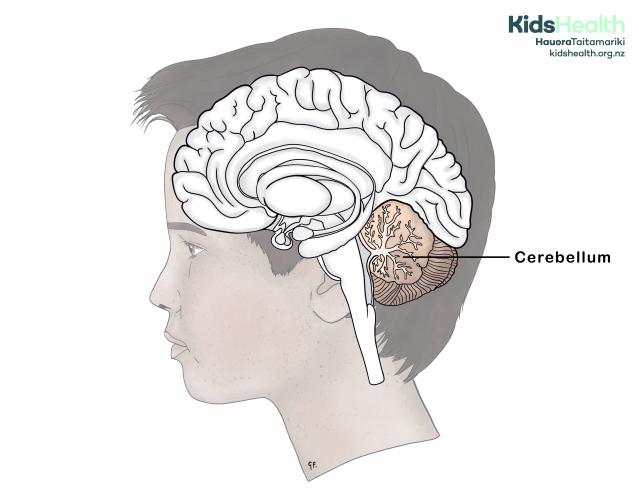
This illustration shows the adolescent brain in cross-section with the cerebellum highlighted.
Source: KidsHealth
transcribeTranscript
A side-view illustration of a young person’s head with the brain shown in cross-section. The cerebellum is shaded brown and located at the lower back part of the brain. A black line points to it with the label 'Cerebellum'.
The rest of the brain is shown in greyscale with detailed folds and structures.
At the top right is the KidsHealth logo with the website: kidshealth.org.nz.
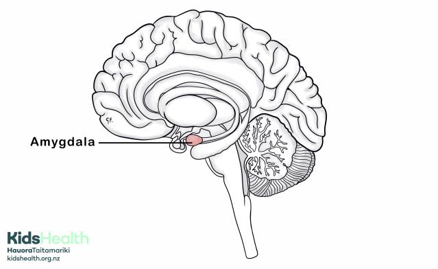
3. The amygdala and hippocampus
Controls emotion and memory.
This illustration shows the adolescent brain in cross-section with the amygdala and hippocampus highlighted.
Source: KidsHealth
transcribeTranscript
A side-view illustration of a young person’s head with the brain shown in cross-section. Two areas in the centre of the brain are coloured and labelled:
- The amygdala is a small circular structure near the front of the brain, shown in light pink and labelled 'Amygdala'.
- The hippocampus is a long, curved structure shown in orange, stretching towards the back of the brain and labelled 'Hippocampus'.
The rest of the brain is drawn in greyscale with detailed folds and internal structures.
At the top right is the KidsHealth logo with the website: kidshealth.org.nz.
4. The cortex
The cortex connects up all the senses and thinking parts. This includes the prefrontal cortex which helps with fine judgement and control.
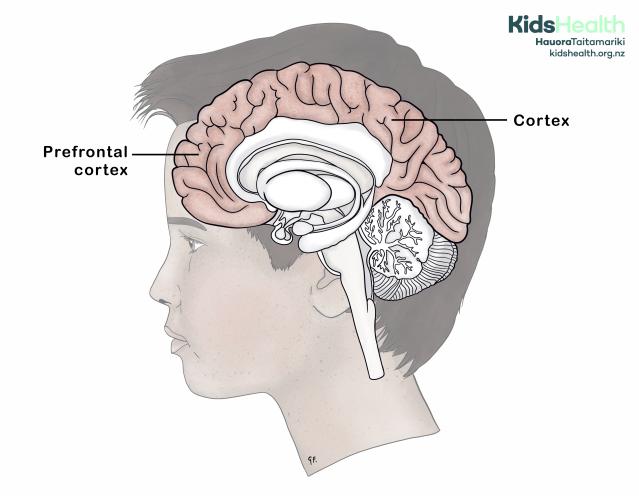
This illustration shows the adolescent brain in cross-section with the cortex and prefrontal cortex highlighted.
Source: KidsHealth
transcribeTranscript
A side-view illustration of a young person’s head with the brain shown in cross-section. The outer layer of the brain is shaded pink to show the cortex, with the front part highlighted more specifically as the prefrontal cortex.
Two black lines point to these areas with the labels:
- 'Prefrontal cortex' – located at the front of the brain.
- 'Cortex' – the outer surface covering the rest of the brain.
The inner parts of the brain are shown in greyscale with detailed folds and internal structures.
At the top right is the KidsHealth logo with the website: kidshealth.org.nz.
Brain development
Scientists believe the brain develops and connects functionally in stages. The emotional areas of the brain (the limbic system) are present at birth. When we're tamariki (children), we are more reliant on parents to help us regulate our emotions. As we grow into teenagers, this becomes more our own job. To do this, new connections form between the thinking and emotional areas of the brain. This also helps us make more adult-level decisions, plans, and thoughts.
Which part of their brain teenagers use most of the time
Rangatahi often 'think with their feelings'
Researchers have done experiments showing that rangatahi often 'think with their feelings'. Researchers have scanned the brain to show which parts light up when a person uses them.
When adults and rangatahi look at faces showing different emotions, the part of their brain that lights up is different.
Adult brain
Adults use their prefrontal cortex to look at faces and try to decide what emotion is happening.
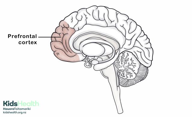
This illustration shows the brain with the prefrontal cortex highlighted.
Source: KidsHealth
transcribeTranscript
A side-view diagram of the brain with the front section shaded pink and labelled 'Prefrontal cortex'. The rest of the brain is drawn in soft greyscale, showing curved brain folds and inner brain structures.
At the bottom left is the KidsHealth logo with the website: kidshealth.org.nz.
Teenage brain
Rangatahi use their amygdala rather than their prefrontal cortex most of the time. In other words, they are using their emotions to try and understand emotion.
This illustration shows the brain with the amygdala highlighted.
Source: KidsHealth
transcribeTranscript
This illustartion shows a side-view diagram of the brain with a small almond-shaped area near the centre coloured pink and labelled 'Amygdala'. The rest of the brain is drawn in soft greyscale, including visible brain folds and structures.
At the bottom left is the KidsHealth logo with the website: kidshealth.org.nz.
It helps if adults can understand how this feels for rangatahi
To understand how this feels, imagine you have lost your keys and you are already late for work. Think about how many times you look for the keys in the same place - maybe 5, 10, or even 20 times. You panic – you no longer think with your cortex, you are thinking with your emotions. Remember how it feels if someone tells you to calm down and think sensibly about when you last had them. That is how your teenager feels when they are running on their emotions. This is because their brain often hasn't developed that linkage.
When talking to rangatahi, be careful to check what emotion they are seeing in you
Often, rangatahi can misinterpret emotions. They may see anger when, in reality, you are feeling anxious. This can often lead to many moments of miscommunication. So, when you are talking to rangatahi, be careful to check what emotion they are seeing in you. Make sure you always acknowledge their emotions first. Then help them to be able to think about what they are feeling.
What can help brain development
Talking to your child really helps
Adults who talk to tamariki as they're growing up really help. Tamariki need a safe environment where they have consistent, loving support. This is vital for the brain to develop well. Rangatahi need adults to believe in them and encourage them.
Rangatahi respond better to rewards than to punishment. They need clear, consistent boundaries. Importantly, parents need to respect their child's growing capacity and independence.
Rangatahi need opportunities to grow many different skills
As they mature and their brains develop, rangatahi need to learn that they don’t have to depend only on their parents. They can build healthy relationships with other trusted adults too. They need opportunities to grow many different skills. They also need to contribute in ways that feel useful and appreciated. The brain develops in a way that produces lots of connections that are then removed if they are not used. So, take care to encourage lots of useful connections.
Develop good habits during the teen years
Another principle is that when connections 'fire together, they wire together'. The teen years are a key time to build good habits, like being active and thinking positively. These habits help shape how the brain works in adulthood.
We know that the brain can change throughout life. But, it is much easier to get the 'wiring' right during childhood than during adulthood.
What harms brain development
Abuse of any sort
It is now well established that any kind of abuse, especially early in life, can affect how a child’s brain develops.
Types of abuse include:
- verbal abuse
- emotional abuse
- physical abuse
- sexual abuse
- neglect
Sometimes, this is hard to change. So it is very important to protect tamariki throughout their development. This is especially important during times of peak brain development. Peak brain development occurs during pregnancy and the first five years of life. The second phase of brain development is around puberty.
Alcohol and drugs
Read about the effect of alcohol and drugs on the developing teenage brain.
Parenting Teens - When Should I Talk To My Child About Drugs?
Parenting Teens - Alcohol & Young People
More information
The Brainwave Trust Aotearoa website provides a range of articles and podcasts about adolescence, including the adolescent brain and the effects of alcohol and marijuana on the adolescent brain.
There are proven steps that you can take to reduce the impact of alcohol on your whānau for now, and for generations to come. Research and advice suggest that delaying drinking is always the best choice for young people. If that doesn't work for your situation, there are other things you can do.
The Drug Foundation has a variety of resources to help parents talk to their teens about alcohol and drugs. Their website has videos and infographics in English, Samoan, Tongan, te reo Māori and Chinese.
Acknowledgements
Illustrations by Dr Greta File. Property of KidsHealth.
Image - Figure 3 from article 'Dynamic mapping of human cortical development during childhood through early adulthood'. Volume 101, issue 21. Copyright 2004. National Academy of Sciences, U.S.A. Our grateful thanks to the National Academy of Sciences, U.S.A. for permission to reproduce the image on this page. Please note this image is copyright and requests to reproduce it elsewhere must be made to the copyright holder.