
Key points about retinopathy of prematurity
- retinopathy of prematurity (ROP) eye exams can be painful for pēpi
- the presence of whānau (family) during these times can reduce stress for both pēpi (babies) and whānau
What is retinopathy of prematurity (ROP)?
Retinopathy of prematurity (ROP) is an eye condition that may affect pēpi who are preterm (born early). Pēpi who are most at risk of ROP are those:
- born less than 30 weeks
- weighing less than 1250 grams at birth
- who have been very unwell, usually in the first month after birth
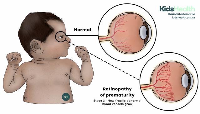
An illustration comparing normal eye anatomy to stage 3 ROP.
Source: KidsHealth
transcribeTranscript
The iIllustration compares normal eye anatomy to stage 3 retinopathy of prematurity (ROP)
On the left is a baby shown facing sideways. Their right eye is circled.
On the right, there are two circular close-ups:
- The top circle is linked to the baby's eye with a solid line. It is labelled Normal. It shows what the inside of a normal eye looks like.
- The bottom circle shows a baby’s eye with stage 3 retinopathy of prematurity. It is labelled Retinopathy of prematurity. Stage 3 - New fragile abnormal blood vessels grow.
At the top right corner is the KidsHealth logo with the website address: kidshealth.org.nz.
A bit about the eye
The retina is at the back of the eye. The retina captures light from our surroundings and sends visual signals to the brain. These become the images we see.
As a baby’s eye develops, tiny blood vessels grow throughout the retina to supply oxygen and nutrients needed for eye development. At 16 weeks of pregnancy, these blood vessels start growing and stop at about one month after birth.

An illustration showing normal eye anatomy.
Source: KidsHealth
transcribeTranscript
The illustration shows the inside of the eye.
The illustration's title is: Looking at the anatomy inside the eye
This illustration shows what the inside of a normal eye looks like.
Labels point to these parts of the eye:
- Cornea
- Pupil
- Lens
- Iris
- Retina
- Retinal blood vessels
- Optic nerve
The label at the bottom left of the illustration: Normal
At the top right is the KidsHealth logo with the website: kidshealth.org.nz.
The cause of ROP
When pēpi are born preterm, the retinal blood vessels may not grow normally, and blood may leak from them. This can cause scarring, and the retina can pull away from the back of the eye, called severe ROP.
ROP can be:
- mild
- moderate
- severe
Mild to moderate ROP
Mild to moderate ROP may not need treatment as over time, the body may grow normal blood vessels in the retina. Mild to moderate ROP is more common than severe ROP.
Severe ROP
Severe ROP often requires treatment to prevent vision loss or permanent blindness.
Why an ROP eye exam is needed
ROP affects the inside of the eye. An eye exam is the only way to diagnose ROP. Regular eye exams are important. If there is a diagnosis of ROP, pēpi can have treatment to prevent vision loss or permanent blindness.
With early treatment, most pēpi gain good or very good eyesight. If your baby is at risk of ROP, they will have an eye exam in your baby's neonatal intensive care unit (NICU). Either an ophthalmologist (eye specialist) will come to the unit, or a neonatal nurse with eye exam training will do the eye exam.
For most pēpi, the first eye exam will happen 4 to 6 weeks after birth.
Before the eye exam
A nurse will give pupil-dilating eye drops to both eyes about 30 to 60 minutes before the eye exam. Pēpi need dilated pupils so the blood vessels of the retina can be seen.
Let the nurse know if you would like to stay for the eye exam and if you would like to have a role during the eye exam. You might want to place a hand on your baby, or karakia (pray) before the eye exam. Or, you might want to leave and come back after the eye exam.
During the eye exam
The eye exam is painful and distressing for pēpi, and pēpi will often cry out. To help ease pain and distress, pēpi will:
- have numbing eye drops
- be swaddled
They can also have:
- expressed breastmilk
- a pacifier
- oral sucrose (sugar)
A small metal device, called a speculum, keeps your baby's eyes open. Most whānau haven't seen an eye test like this before. It can look a bit scary seeing the device holding the eye open with the camera directly on the eye.
A health professional will use a special camera to take photos of the retina. For the camera to work, the health professional will put medical grade jelly on your baby's eye. They will then put the camera directly on your baby’s eyeball.
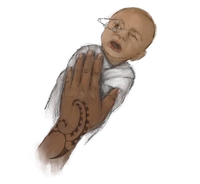
An illustration showing caring for a baby during an eye exam for retinopathy of prematurity (ROP).
Source: Heramaahina Eketone (Ngāti Maniapoto, Waikato)
transcribeTranscript
This illustration is specific for the care given to pēpi during the retinopathy of prematurity eye examination, shown with the handhold touching. Incorporation of moko (tattoo) on the hand represents a combination of care and a gesture of aroha (love), an innovative way to integrate both physical and cultural representations of nurturing. The half-moon cut-outs represent the kākano or seeds to further reinforce the theme of growth. The hand itself is not overly feminine or masculine in appearance, thereby allowing the individual’s gender to be undefined.
The eye exam doesn’t take long to do. Once it is finished, pēpi settle easily.
Having physical touch or singing to your baby during the eye exam may provide comfort for both you and your baby. But, for some whānau, staying for the eye exam is hard. It is OK if you do not want to stay. The nurse will continue to care for your baby.
After the eye exam
As soon as the eye exam is over, talk with your nurse about picking your baby up and giving them a cuddle. You can talk with the ophthalmologist or nurse and ask any questions about the eye exam. You can ask them about the results of the eye exam and when the next eye exam is. Often, pēpi need eye exams every 2 weeks. Some pēpi will need weekly eye exams.
Your baby may have red and puffy eyes, and red marks on their eye lids from the speculum. These skin effects usually disappear within a few hours. They should be gone by the next day.
You might also notice that your baby’s pupils stay dilated for a few hours after the eye exam is over. This means your baby might also be sensitive to light. This is normal, and the pupils should return to normal by the next day.
Treatment for retinopathy of prematurity
Retinopathy Of Prematurity Treatment
Support
For advice and support, call the Parent Helpline on 0800 568 856.
Call the Depression Helpline on 0800 111 757 or free text 4202.