
Key points about congenital cataracts
- newborn pēpi usually have clear lenses inside the eye
- a congenital cataract is cloudiness of the whole lens or part of it, that is present at birth or appears soon after
- the cloudy area can stop light reaching the back of the eye and affect how a baby learns to see
- congenital cataracts can happen in one or both eyes
- early diagnosis and treatment are very important to protect vision
- treatment may include surgery, glasses or contact lenses, and regular eye checks
- pēpi with congenital cataracts often need long-term vision support and monitoring
What are congenital cataracts?
New born pēpi usually have clear lenses inside the eye. A congenital cataract is cloudiness of the whole lens or part of it, that is present at birth or appears soon after.
In a healthy eye, the lens is clear and helps focus light onto the retina, which sends messages to the brain. When the lens is cloudy, light cannot pass through properly. This makes the vision blurry.
Some congenital cataracts are small and do not affect sight. Others are larger or in the centre of the lens and need treatment to prevent permanent vision loss.
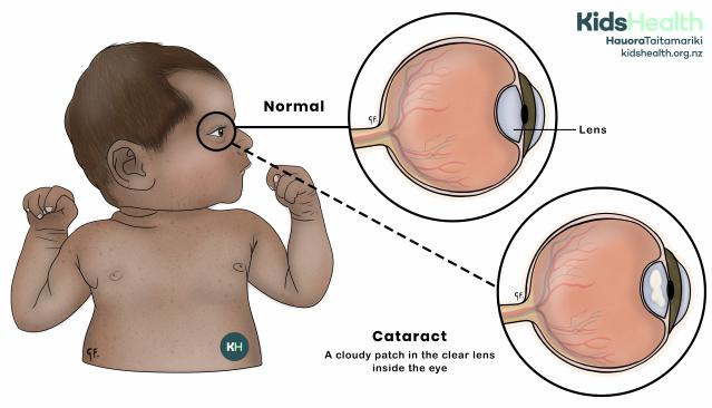
An illustration showing a normal clear lens compared with a cloudy cataract inside the eye.
Source: KidsHealth
transcribeTranscript
The illustration shows a baby on the left, with two enlarged close-ups of the eye on the right.
- The top circle shows the normal eye with a clear lens. The label points to the lens.
- The bottom circle shows a cataract - a cloudy area sits inside the lens.
At the top right of the illustration is the KidsHealth logo with the website: kidshealth.org.nz.
A bit about the eye
The eye has lots of parts including the:
- cornea
- pupil
- lens
- iris
- retina
- optic nerve
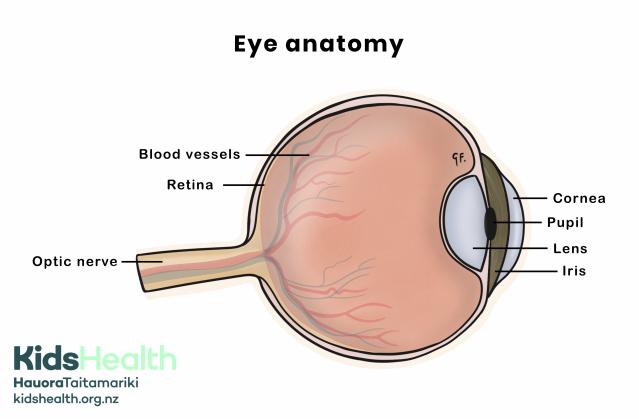
An illustration showing the main parts of the eye, including the retina, blood vessels, optic nerve, cornea, pupil, lens and iris.
Source: KidsHealth
transcribeTranscript
The illustration shows a side view of an eye with the main structures labelled.
- At the back of the eye, the Blood vessels spread over the Retina, and the Optic nerve extends from the back.
- At the front of the eye, the Cornea covers the Pupil, with the Iris around it.
- Behind the pupil sits the clear Lens.
- The illustrations is labelled Eye anatomy.
At the bottom left of the illustration is the KidsHealth logo with the website: kidshealth.org.nz.
Light goes into the eye through the cornea and pupil. The lens helps focus the light onto the retina at the back of the eye. The retina sends messages through the optic nerve to the brain, which makes the picture we see.
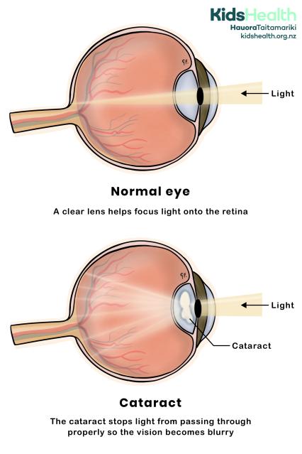
An illustration showing how a normal clear lens focuses light, and how a cataract blocks light and makes vision blurry.
Source: KidsHealth
transcribeTranscript
The illustration shows two side-by-side diagrams of an eye.
- The top diagram is labelled Normal eye and shows a clear Lens that lets light pass straight through to the back of the eye.
- The lower diagram is labelled Cataract and shows a cloudy Cataract in the Lens, which blocks and scatters the light and causes blurry vision.
At the top right of each diagram is the KidsHealth logo with the website: kidshealth.org.nz.
Causes of congenital cataracts
The exact cause of congenital cataracts is not known for many pēpi.
Some possible causes of congenital cataracts include:
- genetic factors – cataracts can run in whānau (families)
- infections during pregnancy – such as rubella or herpes
- syndromes or other health conditions – such as Down syndrome
Symptoms of congenital cataracts
If a baby has a cataract, parents, whānau or health professionals may notice:
- a white or grey cloudy spot in the pupil
- a white glow in photos (instead of the usual red reflection)
- eyes that don’t look in the same direction
- fast or 'shaky' eye movements
- sensitivity to light
- the baby isn’t looking at faces
- the baby isn’t tracking objects that are moving in front of them
Diagnosing congenital cataracts
Newborn and early checks
In Aotearoa New Zealand, all newborn pēpi have an eye check as part of the Well Child Tamariki Ora programme. A health professional will shine a light into the eyes to look for the red reflex (a red glow from the retina).
The health professional will refer your baby for more tests if their reflex is:
- dull
- missing
- uneven between the 2 eyes
Eye specialist assessment
An eye specialist (ophthalmologist) examines the lens and other parts of the eye. The doctor may use eye drops to widen the pupil for a better look inside. The specialist will check if the cataract affects vision. They will discuss whether your baby needs treatment.
Extra tests
Some pēpi need more tests to find out the cause of their cataract. These can include:
- eye photos or scans to measure the cataract
- blood or genetic tests if an inherited or metabolic cause is suspected
- other medical checks if the cataract is possibly part of another condition
Managing congenital cataracts
Treatment depends on how much the cataract affects vision, where it is in the lens, and your child’s age.
Observation
Your child’s ophthalmologist may just arrange regular check-ups if the cataract is:
- small
- off to one side
- not affecting your child's vision
They will watch your child closely.
Surgery
If the cataract blocks vision, surgery is often needed early. This helps the eye and brain learn to see properly. The surgeon removes the cloudy lens through a small opening. In some cases, doctors place an artificial lens in the eye at the same time. This artificial lens is called an intraocular lens or IOL. For very young pēpi, the doctor may wait to add the IOL later and use contact lenses instead.
Your child will need to use eye drops after surgery to prevent infection and inflammation.
Glasses or contact lenses
After surgery, your child will need help to focus light again. Many pēpi wear contact lenses. As your child grows, they may also need glasses. Your child may still need glasses for focusing even if they've had an IOL.
Using an eye patch
Sometimes one eye becomes weaker because the brain relies on the clearer one. The ophthalmologist may suggest patching the stronger eye. Your child may need to wear an eye patch for a certain number of hours each day. This helps the weaker eye work harder and develop stronger vision.
Regular follow-up
Tamariki need regular eye checks after surgery. This is to make sure their vision develops well. Regular checks can also watch for problems like glaucoma or inflammation. The eye team will also adjust glasses or contact lenses as your child’s eyes grow.
Helping your child with congenital cataracts
Medical appointments
Go to all your child’s follow-up appointments. Regular checks allow health professionals to pick up and treat early changes.
Follow your child’s treatment plan
If your child uses contact lenses, clean and replace them as instructed. If your child needs glasses, encourage your child to wear them every day. Consistency is very important for vision development.
Use an eye patch as directed
If the ophthalmologist recommends an eye patch, follow the schedule carefully. An eye patch helps the weaker eye get stronger and supports better vision in both eyes.
Encourage visual play
Offer toys and books with bright, bold colours and different textures. Move toys slowly from side to side to help your baby track movement. Talk to your baby while showing them faces and objects so they start to connect sight and sound.
Support development
Vision affects movement, balance, and learning. Ask your health professional about early intervention services. This includes physiotherapy, occupational therapy, or vision support.
Watch for changes
Notice how your child responds to faces, light, and movement. See a health professional straightaway if your child:
- seems to bump into things
- stops following objects
- has one eye that looks different from the other
- has changes to the way their eyes look
Create a safe and stimulating space
Good lighting helps your child use their vision more easily. Avoid glare from windows or shiny surfaces. Keep furniture and toys in familiar places so your child can move confidently.
More information
Blind Low Vision NZ provides information, community programmes, and family support for people with low vision or blindness. Visit their website to learn more.
BLENNZ offers educational support for children and young people who are blind, low vision, or deafblind. Visit their website for resources and support.
Acknowledgements
Illustrations by Dr Greta File. Property of KidsHealth.