
Key points about developmental hip dysplasia
- developmental dysplasia of the hip (DDH) affects the hip joint in pēpi (babies) and young tamariki (children)
- DDH occurs when the hip joint doesn't form properly, causing instability or dislocation
- early detection and treatment are important for normal hip development
- regular hip checks are part of routine newborn and infant checks
What is developmental hip dysplasia?
Developmental hip dysplasia is a condition where a baby's hip joint doesn't develop correctly. The ball (femoral head) at the top of the thigh bone (femur) may not fit snugly into the hip socket. This means it can come out of the socket (dislocate) easily.
The problem is usually there at birth, but it can also develop during infancy or childhood. DDH can affect one or both hips.
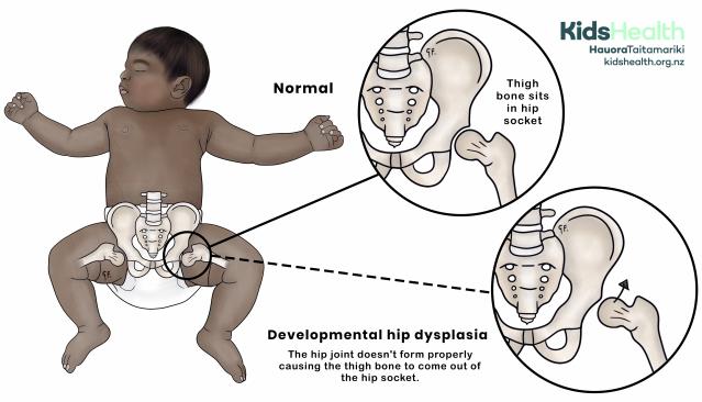
An illustration showing the difference between a normal hip joint and developmental hip dysplasia.
Source: KidsHealth
transcribeTranscript
The illustration shows a baby lying on their back, with legs bent and turned outward. Two circles are connected by dashed lines to the baby’s hip area.
- The top circle is labelled ‘Normal’ and shows the thigh bone sitting correctly in the hip socket.
- The bottom circle is labelled ‘Developmental hip dysplasia’ and shows the thigh bone displaced out of the hip socket. Beneath the image, the text reads: ‘The hip joint doesn’t form properly causing the thigh bone to come out of the hip socket.’
At the top right is the KidsHealth logo with the website kidshealth.org.nz
A bit about the hip
The hip joint includes the:
- thigh bone (femur)
- hip bone (pelvis)
- muscles, ligaments and tendons around the hip joint act like strong ropes to hold the bones together
The hip joint works like a ball and socket joint. The top of the thigh bone is shaped like a ball which is called the femoral head. The femoral head sits inside a cup-shaped socket in the pelvis.
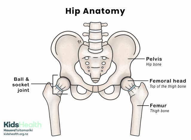
An illustration of normal hip anatomy showing the pelvis (hip bone), femur (thigh bone), and how the top of the thigh bone fits into the hip socket to form the ball and socket joint.
Source: KidsHealth
transcribeTranscript
The illustration shows a front view of the pelvis and upper thigh bones. Each hip joint is shown where the top of the thigh bone fits into the hip socket.
- The left side of the image is labelled Ball & socket joint where the thigh bone meets the pelvis.
- The top of the thigh bone is labelled Femoral head – Top of the thigh bone.
- The hip bone is labelled Pelvis – Hip bone.
- The long thigh bone is labelled Femur – Thigh bone.
- The illustration is titled Hip Anatomy.
At the bottom left is the KidsHealth logo with the website: kidshealth.org.nz.
Causes of developmental hip dysplasia
The exact cause of DDH is not known.
DDH can affect any baby. It is more common in female pēpi. Other things that may give a baby a higher chance of having DDH include:
- having a whānau (family) member with hip dysplasia
- being the firstborn child
- being in a breech (feet first) position during birth
- low fluid volumes during pregnancy (oligohydramnios)
Signs and symptoms of developmental hip dysplasia
Common signs and symptoms of DDH include:
- 1 leg looking shorter than the other
- uneven skin folds on the thighs or buttocks
- reduced movement or flexibility in 1 leg
- a clicking felt in the hip when changing your baby’s nappy or dressing them
- a limp when your child starts walking
Many pēpi and tamariki with DDH might not show obvious signs.
Diagnosing developmental hip dysplasia
Examining the hips
Your baby will have their hips checked during their newborn check. Your midwife, doctor and Plunket nurse will examine your baby for hip problems several times in the first months of life. This involves some gentle movement tests to check your baby’s hip stability.
Scan
If a health professional thinks your baby may have hip dysplasia, they will arrange an ultrasound scan of your baby’s hips. Pēpi and tamariki over 6 months will have an x-ray.
If you have a baby who is at higher risk of DDH, you may be offered an ultrasound scan of their hips around 6 weeks of age.
Managing developmental hip dysplasia
Treatment for DDH depends on how old your baby is and how badly their hip or hips are affected.
If your baby has DDH, they will see an orthopaedic surgeon (a doctor who specialises in bones and joints). The orthopaedic surgeon will examine your child and look at their scans then plan the best treatment for them.
Harness or brace
A harness or brace is used for younger pēpi diagnosed with DDH. A hip harness or brace can help keep the hip joint in the right position to help it develop normally.
Different types of harnesses or braces can be used, including a Pavlik harness and an abduction brace (rhino cruiser).
See the information booklets from Starship for more information on the Pavlik harness and abduction brace.
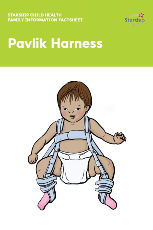
Visit the Starship website to see their information booklet on the Pavlik harness.
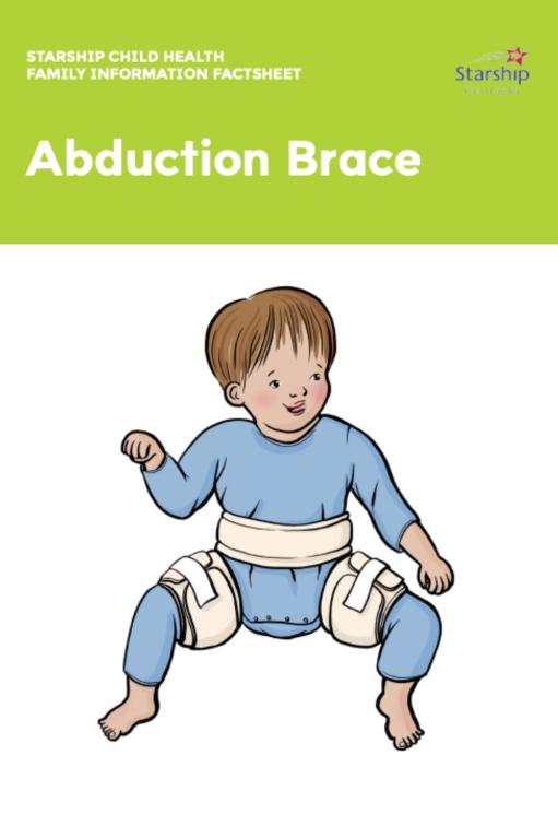
Visit the Starship website to see their information booklet on the abduction brace.
Surgery
Pēpi with more severe DDH or those who are older when diagnosed may need surgery. There are different procedures for treating DDH.
Closed reduction procedure
Closed reduction is when your child's hip joint is moved into the right position while they are asleep under general anaesthetic. This is usually done with dye injected into the hip joint (arthrogram) and often the tight tendon in the groin is released through a small cut.
Open reduction procedure
Open reduction is done when your child’s hip joint is unable to be safely placed in the correct position while they are asleep under general anaesthetic. Surgery is usually through a cut in their groin. The hip is placed into the correct position after releasing the soft tissues that are holding the hip out of joint.
In older tamariki, this can be done at the same time as bony (osteotomy) procedures to realign the pelvis (socket) or femur (ball).
Osteotomy
Occasionally tamariki continue to have a steep socket or the ball does not sit snugly under the cup. In these cases, your surgeon may recommend an osteotomy (bony) procedure. This is when more surgery is done on the thigh or pelvic bone to make sure the hip joint stays in place and develops normally. Surgery in childhood can help prevent your child from developing arthritis in their hip as a young adult.
Hip Spica
After closed or open reduction surgery, your child will usually be placed in a hip spica. This is a cast that covers your child’s body from their waist down to their knees. Some tamariki need hip spicas for several months.
See the information booklet from Starship for more information on hip spicas.
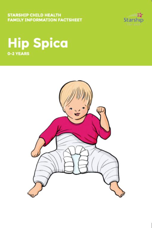
Visit the Starship website to see their information booklet on the hip spica for 0 to 2 year olds.
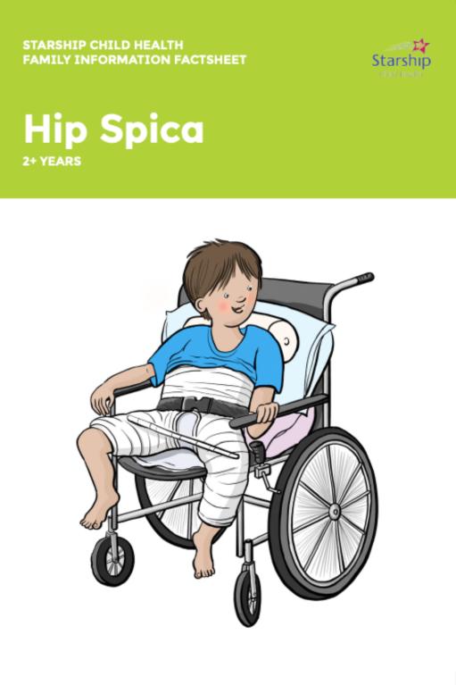
Visit the Starship website to see their information booklet on the hip spica over 2 year olds.
Preventing developmental hip dysplasia
How your baby’s legs are positioned during the first few months of their life may affect their chance of developing DDH.
Using safe swaddling techniques is important. Your baby’s legs should always be able to bend and move freely. Baby carriers and slings that support the thighs and allow the legs to spread in an “M” or “frog-leg” position help support healthy hip development.
Regular hip checks by health professionals during baby check-ups help catch any problems early. If you have a family history of DDH or your baby was breech, extra monitoring may be needed.
What to expect long-term
With early treatment, most tamariki with DDH will have normal hip development with no long-term issues. Regular follow-ups ensure the hip joint is developing properly.
If DDH is not treated or picked up later in life, it may be more challenging to treat. Some tamariki and adults may develop hip pain and arthritis when they’re older.
Acknowledgements
Illustrations by Dr Greta File. Property of KidsHealth.