

Key points about urinary tract dilatation
- urinary tract dilatation (UTD) is often found during a routine pregnancy ultrasound scan
- UTD is something seen on a scan, not a disease or illness by itself
- there is nothing you did to cause UTD and nothing you could have done to prevent it
- in most pēpi, UTD improves before or after birth
- your baby may need follow-up scans, even if they seem well
- some pēpi need more tests or ongoing check-ups
- only a small number of pēpi need treatment or surgery
The urinary tract and how it works
The urinary tract includes the kidneys, ureters, bladder, and urethra.
The kidneys filter and remove waste and water from the blood to produce wee (urine). The wee travels from the kidneys down 2 narrow tubes called the ureters. Wee is then stored in the bladder.
When your baby does a wee, urine flows out of the body through the urethra, a tube at the bottom of the bladder. The opening of the urethra is at the end of the penis in boys and in front of the vagina in girls.
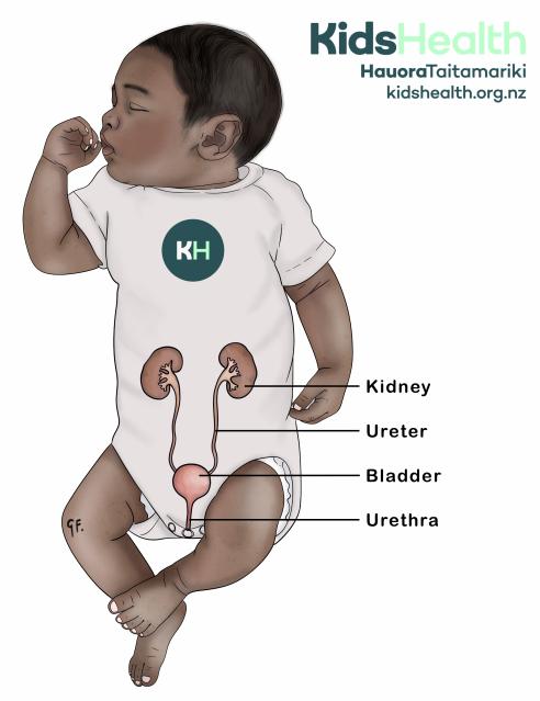
This illustration shows the urinary system in a baby.
Source: KidsHealth
transcribeTranscript
The illustration shows a baby wearing a white bodysuit, with an overlay of the urinary system on their body.
The urinary system includes:
- 2 kidneys
- 2 ureters, which run from the kidneys to the bladder
- the bladder
- the urethra, which carries urine out of the bladder
The kidney, ureter, bladder and urethra are labelled.
At the top right is the KidsHealth logo with the website: kidshealth.org.nz.
What is urinary tract dilatation?
Urinary tract dilatation means part of the urinary tract looks wider than expected on an ultrasound scan. UTD is a scan finding. It does not tell the healthcare team what is causing the widening.
The part of the kidney that collects urine is called the renal pelvis. In UTD, the renal pelvis or another part of the urinary tract looks wider than usual.
UTD can affect one or both kidneys.
When health professionals find UTD before birth, they call it antenatal UTD. If it continues after birth, they call it postnatal UTD.
You may also hear UTD referred to as renal dilatation or hydronephrosis.
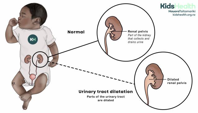
This illustration compares a normal renal pelvis with a dilated renal pelvis in urinary tract dilatation.
Source: KidsHealth
transcribeTranscript
The illustration shows a baby with an overlay of the kidneys, ureters, bladder and urethra on their body. A circle highlights one kidney, with 2 enlarged diagrams comparing a normal renal pelvis with a dilated renal pelvis.
- The top diagram is labelled Normal. It shows a kidney and ureter. The renal pelvis is labelled. Text explains that the renal pelvis is the part of the kidney that collects and drains urine.
- The bottom diagram is labelled Urinary tract dilatation. It shows the renal pelvis looking wider than in the normal kidney. The widened area is labelled Dilated renal pelvis.
- Text below the bottom diagram reads: Urinary tract dilatation. Parts of the urinary tract are dilated.
At the top right is the KidsHealth logo with the website: kidshealth.org.nz.
Causes of urinary tract dilatation
There is nothing you did to cause UTD and nothing you could have done to prevent it.
In many pēpi, UTD is temporary. It often improves before birth or shortly after. Health professionals often do not find a specific cause.
UTD can be linked with:
- a narrowing or blockage that affects how urine drains
- vesicoureteric reflux (VUR), where urine flows backwards from the bladder towards the kidneys
- differences in the structure of the kidneys, ureters or bladder
- a duplex collecting system, where 2 ureters drain one kidney
UTD usually happens on its own. Most pēpi do not have differences in other parts of their body.
If a scan shows other differences, your baby’s healthcare team will discuss these with you. They may offer further testing.
Finding urinary tract dilatation
Health professionals usually find UTD during a routine pregnancy ultrasound scan.
The scan checks:
- your baby’s kidneys and bladder
- the tubes that drain urine
- how much fluid is around your baby
- your baby’s growth and wellbeing
Your baby’s healthcare team will check how much dilatation there is and whether it affects one or both kidneys. They use the scan findings to give the UTD a grade. The grade helps them plan your baby’s follow-up.
It is not always possible to find the cause before your baby is born. If the dilatation remains after birth, your baby may need more scans and tests.
Urinary Tract Dilatation Grades & Follow-Up
Symptoms of urinary tract dilatation
UTD does not cause symptoms before birth.
Most pēpi with UTD remain well after birth. UTD itself does not usually make pēpi look or act unwell.
Some pēpi with UTD have a higher chance of developing a urinary tract infection (UTI). Your baby’s healthcare team will explain what signs to look out for.
Managing urinary tract dilatation
During pregnancy
Your baby’s healthcare team may arrange more ultrasound scans. The timing depends on what the scans show.
Mild dilatation often improves before birth or remains mild. More significant dilatation needs closer checking. Your healthcare team may refer you to a maternal fetal medicine service. This service specialises in caring for pēpi before birth.
Care during pregnancy usually involves scans and planning for after your baby is born.
UTD on its own does not usually affect how or where you give birth. In most cases, you can give birth at your local hospital.
Some pēpi need specialist care after birth. Your healthcare team may recommend that you give birth at a hospital that can provide this care.
After birth
Whether your baby needs an ultrasound after birth, and when they have it, depends on the pregnancy scan findings.
Your baby’s healthcare team will tell you when your baby needs their scan. Some pēpi need a scan soon after birth. Others have their first scan when they are a little older.
Take your baby to all follow-up appointments, even if they seem completely well. Scans allow the healthcare team to check whether the dilatation is improving.
Tests your baby may need
Not all pēpi need further tests. It depends on what their scans show.
Your baby may need:
- another kidney and bladder ultrasound
- a bladder x-ray test called an MCUG, which checks whether urine flows back towards the kidneys
- a nuclear medicine kidney scan, which checks how well the kidneys work and how well urine drains
- urine tests to check for infection in the wee
- blood tests to check how well the kidneys are working
Micturating Cysto-Urethrogram In Children
Nuclear Medicine Kidney Scans In Children
Treatment for urinary tract dilatation
Most pēpi do not need treatment. They may still need regular ultrasound scans to check the dilatation.
Treatment depends on the cause and the scan findings. Your baby may need:
- a small daily dose of an antibiotic to help prevent urinary tract infections
- surgery to relieve a blockage and help urine drain
Only a small number of pēpi need surgery.
Arranging your baby’s follow-up
Your maternity team, such as your midwife or obstetric team, usually arranges your baby’s first ultrasound after birth.
Enrol your baby at a general practice clinic soon after birth. The health professionals involved in your baby’s care will help make sure your baby has the scans and appointments they need.
If you have not received information about your baby’s next scan, contact your baby’s healthcare team. It is fine to check.
Tell your baby’s healthcare team if you change your phone number or move house so they can continue to contact you.
Long-term outlook for urinary tract dilatation
The outlook for most pēpi with UTD is very good. Most cases improve on their own before birth or shortly after.
Even when UTD remains, many pēpi do not have an underlying condition that needs treatment. The dilatation may stay the same or slowly improve over time.
Some tamariki (children) need ongoing scans or specialist follow-up. A small number need surgery to improve urine drainage and protect their kidneys.
Very severe UTD can sometimes affect the growth or function of a kidney. If one kidney is affected, the other kidney can often do the work of both. Your child may need ongoing checks of their kidney function and blood pressure.
Follow-up helps the healthcare team identify which tamariki need treatment and which can stop having scans.
Acknowledgements
Illustrations by Dr Greta File. Property of KidsHealth.
